home contact company.overview news sitemap print
![]()
![]() Electrophysiology
Electrophysiology
![]()
Contact |
|
Dr. Timm Danker |
|

 +49 7121 51530 896
 Zeiss Cell Explorer
 Hamamatsu FDSS/uCell
Imaging
Besides classical electrophysiological approaches we offer life cell imaging based techniques in order to monitor cell-cell interactions / analyze compound induced effects on intracellular ion homeostasis.
Monitoring the dynamic changes of the intracellular Ca2+ concentration enables the understanding of cardiac function including inotropic effects and the occurrence of proarrhythmic events.
Two technologies are available for detailled investigations of the intraclellular Ca2+ concentrations:
- Zeiss Cell Explorer with attached Yokogawa spinning disk for rapid confocal imaging offers subcellular resolution with sampling frequencies up to 80 Hz
- Hamamatsu FDSS/Cell is our higher throughput working horse with parallel recordings in 96- or 384-well format at a sampling frequency of up to 120 Hz.
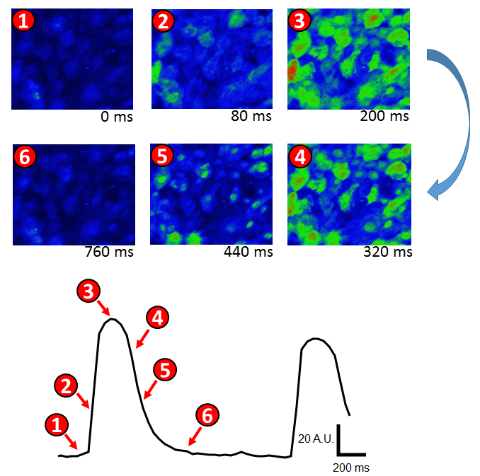
Confocal Ca2+ imaging of cultivated, spontaneously beating cardiomyocytes using the Zeiss Cell Observer system. Top: Time series of false-color coded Ca2+ dependent fluorescence of Cal520-AM loaded cardiomyocytes. Excitation leads to an transient intracellular Ca2+ increase, visualized by a color shift from blue via green to red. Bottom: Fluorescence ntensity-over time trace of above shown recording. Numbers indicate time points of the images.
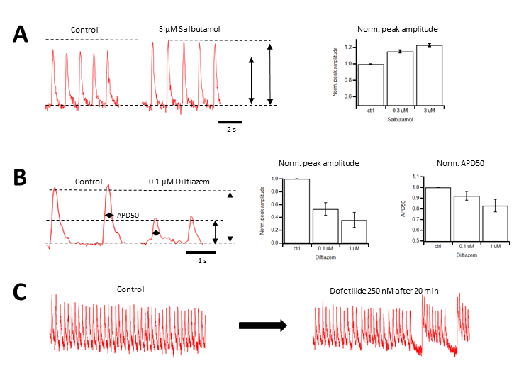
Intracellular Ca2+ Imaging of spontaneously beating cardiomyocytes using the higher throuput Hamamatsu FDSS/uCell system. A: Positive inotropic effect by Salbutamol. Application of this beta2-sympathomimetic drug results in a concentration-dependent increase of intracellular Ca2+ concentration during each beat. B: The Ca2+ channel Inhibitor Diltiazem both reduces Ca2+ transient amplitude and duration,, resulting in negative inotropicity. C: Application of the potent hERG channel inhibitor Dofetilide results in the detection of proarrhythmic behavior in the investigated cardiomyocytes.
